Activating the Clot, Hemolysis, and the Art of Preanalytics
The Scoop - No.4, April 2026 - A Greiner Bio-One Newsletter
Introducing: Inside the tube - Serum Edition
As a reminder, our Inside the Tube series is your go‑to guide for understanding the science behind the blood collection tubes we use every day. Each edition highlights a different tube type, exploring its additives, purpose, and role in laboratory testing while following the recommended order of draw.
This edition will look into the next tube collected: Serum.
The Order of Draw:
1. Blood Culture Bottles

2. Coagulation Tubes

3. Serum Tubes
Serum tubes contain clot activators that can trigger clotting if they contaminate other samples. Drawing them after coagulation tubes prevents additive carryover that could compromise sensitive tests like PT or a PTT.

4. Heparin Tubes

5. EDTA Tubes

6. Glycolytic Inhibitor Tubes
Activating the clot, protecting the results
WHY IS THIS TUBE DRAWN SECOND?
Serum tubes are drawn after coagulation tubes to prevent additive carryover.
For more information, refer to The Scoop, Edition 2: “Sequence for Success.”
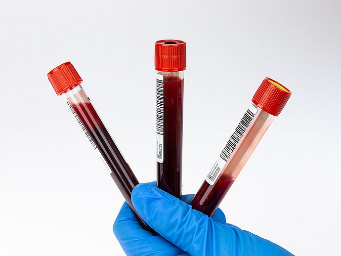
INSIDE THE TUBE
Additive
Serum Tubes are coated with micronized silica particles which activate clotting when tubes are inverted.1, 2
What is a Clot Activator
Clot activators are substances that promote blood clotting by stimulating the coagulation cascade.3 In serum tubes, a commonly used clot activator is micronized silica, a finely powdered form of silicon dioxide. Silica triggers the intrinsic pathway of the coagulation cascade by activating factor XII upon contact with blood.4
How does it work
When blood is drawn into a serum tube containing micronized silica, and gently inverted, the clot activator helps to initiate and accelerate clot formation. Clotting typically occurs within 30 minutes.
WHY PROPER HANDLING IS IMPORTANT
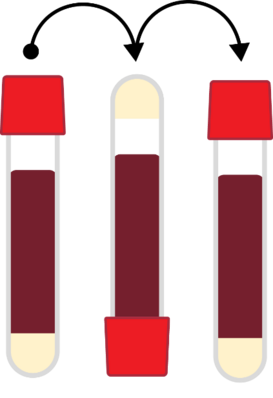
Proper handling of serum tubes is critical for accurate results. Key steps include:
- Adequate mixing (gentle inversions):
to allow the sample to adequately mix with the clot activator additive. - Sufficient clotting time:
at least 30 minutes in an upright position. Note: Tubes containing thrombin-based clot activators reduce the clotting time to approximately 5 minutes. - Timely and correct centrifugation:
within 2 hours of collection.
NOTE
Improper handling like insufficient inversions, premature centrifugation, or temperature extremes, can lead to incomplete clotting, fibrin strands, or hemolysis.5 These preanalytical errors can compromise sample quality and can cause delays, redraws, or erroneous results.
Sources:
1. Greiner Bio-One North America Inc. (2020). VACUETTE® Blood Collection Tubes: Instructions for Use (Rev. 09, 980200B_ Rev09_02-2020).
2. Greiner Bio-One GmbH. (2022). VACUETTE® Blood Collection Tubes: Instructions for Use (Rev. 24, 980200_Rev24_05-2022).
3. Bowen, R. A., & Remaley, A. T. (2014). Interferences from blood collection tube components on clinical chemistry assays. Biochemia Medica, 24(1), 31–44. doi.org/10.11613/BM.2014.005.
4. Lima-Oliveira, G., Lippi, G., Salvagno, G. L., Montagnana, M., & Plebani, M. (2013). Clot activators and anticoagulant additives for blood collection. Biochemia Medica, 23(3), 265–276. doi.org/10.11613/BM.2013.031.
5. Lippi, G., & Plebani, M. (2012). Preanalytical phase—a continuous challenge for laboratory professionals. Clinical Chemistry and Laboratory Medicine, 50(7), 1115–1126.
The Art of Preanalytics: Essentials for Success with Serum Gel Tubes

In May of 2025, Greiner Bio-One hosted an educational webinar titled, “The Art of Preanalytics – Essentials for Success with Serum Gel Tubes”, presented by Sandra de Kozlowski, Scientific Affairs Manager at Greiner Bio-One.
The webinar provides practical insights into the proper handling and use of serum gel tubes (SGTs). This highly informative session covered:
- The evolution and design of serum gel tubes
- Preanalytical best practices, including order of draw, proper inversions and clotting time
- Common handling errors and their impact on sample quality
- Post-centrifugation considerations for aliquoting, transport, and storage
For those who missed it or would like a refresher, the webinar is available on demand:
Watch Now: The Art of Preanalytics - Essentials for Success with Serum Gel Tubes


Hemolysis on Greiner Bio-One Talks
Our podcast Greiner Bio-One Talks dives into hemolysis in the preanalytical phase with two lively episodes. Dr. Cradic and Prof. Salvagno share practical tips for minimizing hemolysis, drawing on their experiences in the U.S. and Italy.
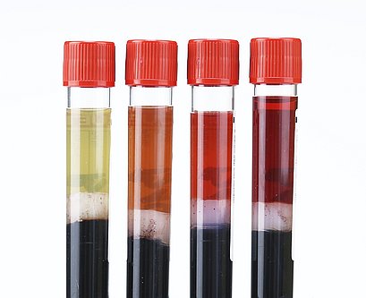
Hemolysis happens but it's preventable
Breaking it Down, literally
Let’s talk hemolysis. The word comes from Latin: hemo = blood and lysis = break apart. It simply means “breaking open blood cells”.
When red blood cells (also called erythrocytes) get damaged, they burst open. This releases hemoglobin (the protein that carries oxygen) and other components into the surrounding plasma (the liquid part of blood). It’s like popping a balloon and everything inside spills out!
Why hemolysis matters:
Hemolyzed samples can lead to misdiagnosis, delayed treatment, and unnecessary costs. One study estimated that in an Emergency Department with ~100,000 visits per year and a 10% hemolysis rate, the cost of hemolyzed samples could hit $4 million annually.1
Top Hemolysis Culprits:
- Prolonged tourniquet time2
- Using an inappropriate needle gauge3
- “Digging” for veins3
- Syringe collection4
- Vigorously shaking/inverting tubes3
- Centrifuging for prolonged speeds7
- Re-centrifuging samples5
- Rough pneumatic tube transport7
- Exposure to high temperatures during storage or transport8,9
Sources:
1. Phelan MP, Ramos C, Walker LE, Richland G, Reineks EZ. The hidden cost of hemolyzed blood samples in the emergency department. J Appl Lab Med. 2021;6(6):1607-1610. doi:10.1093/jalm/jfab086.
2. Saleem S, Mani V, Chadwick MA, Creanor S, Ayling RM. A prospective study of causes of haemolysis during venepuncture: tourniquet time should be kept to a minimum. Ann Clin Biochem. 2009;46(3):244-246. doi:10.1258/acb.2009.008228. PMID:19389888.
3. Wan Azman WN, Omar J, Koon TS, Tuan Ismail TS. Hemolyzed specimens: major challenge for identifying and rejecting specimens in clinical laboratories. Oman Med J. 2019;34(2):94-98. doi:10.5001/omj.2019.19. PMID:30918601; PMCID:PMC6425048.
4. Carraro P, Servidio G, Plebani M. Hemolyzed specimens: a reason for rejection or a clinical challenge? Clin Chem. 2000;46(2):306- 307. PMID:10657399.
5. CLSI. Handling, Transport, Processing, and Storage of Blood Specimens for Routine Laboratory Examinations. 1st ed. CLSI guideline PRE04. Clinical and Laboratory Standards Institute; 2023.
6. Lippi G, von Meyer A, Cadamuro J, Simundic AM. Blood sample quality. Diagnosis. 2019;6(1):25-31. doi:10.1515/dx-2018-0039.
7. Kara H, Bayir A, Ak A, et al. Hemolysis associated with pneumatic tube system transport for blood samples. Pak J Med Sci. 2014;30(1):50-58. doi:10.12669/pjms.301.4228. PMID:24639830.
8. Utoh J, Harasaki H. Damage to erythrocytes from long-term heat stress. Clin Sci (Lond). 1992;82(1):9-11. doi:10.1042/cs0820009.
9. Cadamuro J, Mrazek C, Leichtle AB, et al. Influence of centrifugation conditions on the results of 77 routine clinical chemistry analytes using standard vacuum blood collection tubes and the new BD-Barricor tubes. Biochem Med (Zagreb). 2018;28(1):010704. doi:10.11613/BM.2018.010704. PMID:29187797; PMCID:PMC5701775.
Additional Sources:
1. Simundic AM, Baird G, Cadamuro J, Costelloe SJ, Lippi G. Managing hemolyzed samples in clinical laboratories. Crit Rev Clin Lab Sci. 2020 Jan;57(1):1-21. doi: 10.1080/10408363.2019.1664391. Epub 2019 Oct 11. PMID: 31603708.
2. Šimundić AM, Dukic L, Radisic Biljak V. Preanalytical variation and preexamination processes. Tietz Textbook of Clinical Chemistry and Molecular Diagnostics, ed 7, edited by Nader Rifai, Rita Horvath, and Carl Wittwer, St. Louis, Missouri, Elsevier, 2022; p:80-129.

