Stabilizing Glucose, Honoring the Order, and Saluting a Pioneer
The Scoop - No.2, June 2025 - A Greiner Bio-One Newsletter
When collecting blood samples for glucose testing, proper handling is essential to ensure accurate and reliable results. A critical step in this process is to either place the blood tube on ice immediately or centrifuge it as soon as possible. But why is this necessary?
After blood is drawn, the cells in the sample—especially red and white blood cells—remain metabolically active. These cells continue to consume glucose through a process called glycolysis, converting it into energy. If the sample isn’t processed promptly, glycolysis will continue, causing the glucose concentration in the sample to drop, leading to falsely low results. In fact, studies have shown that glucose levels in unprocessed blood samples can decrease by 5–7% per hour at room temperature - a significant concern when screening or monitoring for conditions such as diabetes or hypoglycemia [1].
Although additives like sodium fluoride are commonly used to inhibit glycolysis, their effect is not immediate. Glycolysis can still occur during the first hour after collection, even in fluoride-containing tubes.
TO PREVENT THIS GLUCOSE DEGRADATION:
- Placing the tube on ice rapidly cools the sample, slowing down the metabolic activity of the blood cells and delaying glycolysis [1], [2].
- Immediate centrifugation separates the plasma or serum from the blood cells, effectively stopping further glucose consumption [1].
An alternative, and increasingly recommended option by the American Diabetes Association, is the use of specialized blood collection tubes that contain a citrate buffer, sodium fluoride, and EDTA [1] - such as the VACUETTE® FC Mix Tube. This additive combination works immediately to inhibit glycolytic enzyme activity, offering a more effective and reliable method of glucose stabilization [1]. Unlike standard tubes, these do not require icing or immediate centrifugation.
SOURCES:
-
David B. Sacks, Mark Arnold, George L. Bakris, David E. Bruns, Andrea R. Horvath, Ake Lernmark, Boyd E. Metzger, David M. Nathan, M. Sue Kirkman; Guidelines and Recommendations for Laboratory Analysis in the Diagnosis and Management of Diabetes Mellitus. Diabetes Care 1 October 2023; 46 (10): e151-e199.
-
CLSI. Handling, Transport, Processing, and Storage of Blood Specimens for Routine Laboratory Examinations. 1st ed. CLSI guideline PRE04. Clinical and Laboratory Standards Institute; 2023.
SEQUENCE FOR SUCCESS
The order of draw during blood collection is a critical component of phlebotomy that directly impacts the accuracy and reliability of laboratory test results. When multiple blood specimens are collected during a single venipuncture, the sequence in which the collection tubes are filled plays a vital role in preventing cross-contamination of additives, which can compromise test outcomes.
Greiner Bio-One follows the order of draw recommendations outlined by CLSI, WHO, and EFLM [1], [2], [3], [4]:
1. Blood Culture Bottles
are drawn first to avoid contamination. Drawing them later could allow additives from other tubes to enter the culture bottles, potentially inhibiting bacterial growth and leading to false-negative results.

2. Coagulation Tubes
are collected second. These tubes are highly sensitive to contamination from other additives such as EDTA, which can chelate calcium - an essential component in the coagulation cascade - resulting in falsely prolonged clotting times in tests like prothrombin time (PT) and activated partial thromboplastin time (aPTT) [4]. Serum tubes with clot activators can also interfere with coagulation testing and should not precede coagulation tubes.
Learn more

3. Serum Tubes
are drawn next to prevent contamination that could hinder clot formation. Serum tubes contain a clot activator.
Learn more

4. Heparin Tubes
are collected after serum. Heparin acts as an anticoagulant by activating antithrombin which blocks the coagulation cascade. Drawing heparin tubes before serum could result in cross contamination and interfere with the clot formation in serum samples.
Learn more

5. EDTA Tubes
which are collected after serum and heparin due to their potential to significantly alter chemistry test results. EDTA contamination can falsely elevate potassium levels and decrease calcium, magnesium, and alkaline phosphatase measurements.
Learn more

6. Glycolytic Inhibitor Tubes
(e.g., those containing sodium fluoride and potassium oxalate) are drawn last. These additives can interfere with potassium and calcium levels, and oxalate may impact coagulation tests by binding calcium.
Learn more
7. Other Additives
please refer to your country-specific Instructions for Use (IFU) for guidance on the order of draw for other additives.
NOTE
If a winged blood collection set is used, the first tube in the series will be underfilled. Therefore, if a coagulation specimen is drawn first, a discard tube (no additive) is recommended prior to collection to ensure the proper additive-to-blood ratio.
Additionally, it is important to recognize that modern plastic red-top tubes are not true “no-additive” tubes. While older glass red-tops contained no additives, current plastic versions typically include a clot activator.
SOURCES
- CLSI. Collection of Diagnostic Venous Blood Specimens. 8th ed. CLSI standard PRE02. Clinical and Laboratory Standards Institute; 2025
- WHO guidelines on drawing blood: best practices in phlebotomy (2010).
- EFLM-COLABIOCLI Recommendation for venous blood sampling, Simundic et al on behalf of the WG-PRE, EFLM and WG-PRE-LATAM of COLABIOCLI. Clin Chem Lab Med 2018 doi 10.1515/cclm-2018-0602.
- Cornes M, van Dongen-Lases E, Grankvist K, Ibarz M, Kristensen G, Lippi G, Nybo M, Simundic AM; Working Group for Preanalytical Phase (WG-PRE), European Federation of Clinical Chemistry and Laboratory Medicine (EFLM). Order of blood draw: Opinion Paper by the European Federation for Clinical Chemistry and Laboratory Medicine (EFLM) Working Group for the Preanalytical Phase (WG-PRE). Clin Chem Lab Med. 2017 Jan 1;55(1):27-31. doi: 10.1515/cclm-2016-0426. PMID: 27444170.
- Banković Radovanović P, Živković Mikulčić T, Simović Medica J. Unexpected abnormal coagulation test results in a 2-year-old child: A case report. Biochem Med (Zagreb). 2020;30:011002.
- Bazzano, G.; Galazzi, A.; Giusti, G.D.; Panigada, M.; Laquintana, D. The Order of Draw during Blood Collection: A Systematic Literature Review. Int. J. Environ. Res. Public Health 2021, 18, 1568
Honoring Dr. Davide Grenache: A Preanalytical Legacy

As 2024 came to a close, the laboratory medicine community bid farewell to one of its most respected leaders. Dr. David Grenache, who retired in December, leaves behind an impressive 35-year legacy of scientific leadership, innovation, and dedication to advancing diagnostic medicine.
Dr. Grenache served as the Chief Scientific Officer for TriCore Reference Laboratories in Albuquerque, New Mexico, where he also led the TriCore Research Institute. In that role, he played a pivotal part in driving innovation in healthcare through clinical device trials, advanced central lab services, and the development of a robust sample biorepository.
Beyond his leadership at TriCore, Dr. Grenache held multiple key roles including laboratory director of TriCore’s flagship core lab, medical director of immunology and point-of-care testing, and as well as clinical professor of pathology at the University of New Mexico. He also served as President of the Association for Diagnostics and Laboratory Medicine (formerly AACC), further shaping the future of the field at a national level.
A PhD graduate in biomedical sciences from Worcester Polytechnic Institute, Dr. Grenache completed his postdoctoral training in clinical chemistry at Washington University School of Medicine in St. Louis. Earlier in his career, he served as an assistant professor of pathology and laboratory medicine at the University of North Carolina at Chapel Hill, as well as a clinical professor of pathology at the University of Utah and ARUP Laboratories.
With more than 150 published articles, abstracts, and book chapters, he is widely recognized for his expertise, particularly in the diagnostic management of pregnant patients and the use of longitudinal lab data to assess individual and population health.
HEAR FROM DR. GRENACHE ON GREINER BIO-ONE TALKS
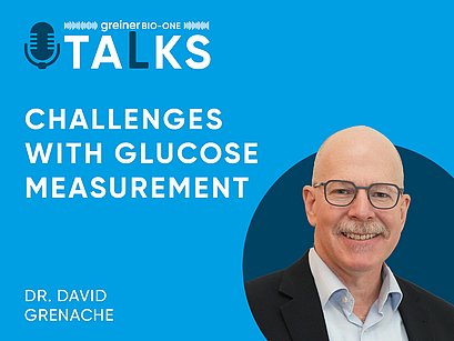
As part of our farewell tribute, we’re thrilled to share our recent podcast episode featuring Dr. Grenache. In this conversation, he offers expert insights into glucose stabilization methods—discussing current practices, their effectiveness, and what the future might hold.
Please join us in thanking Dr. Grenache for his tremendous contributions to laboratory medicine and in celebrating the lasting impact he’s made on the field.

